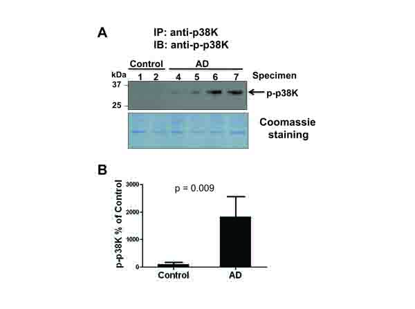
Fig. (2)
Activation of p38K in AD individuals. (A) To accurately determine the activation status of phospho-p38K, brain cytosolic proteins (500 μg/analysis) from AD and control individuals were used for immunoprecipitation (IP) with the specific antibody against p38K. Immunoprecipitated proteins were separated on 12% SDS-PAGE gel, transferred to PVDF-Immobilon, and subjected to IB analysis using an antibody against phospho-p38K (top panel) or Coomassie blue stained (bottom panel). Longer exposure of the same blot did not show any phospho-p38K band in control subjects. (B) Densitometric analysis of the immunoblot shows a marked increase in phospho-p38K in AD individuals. P=0.009.

