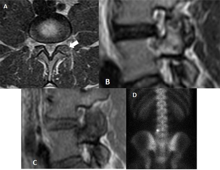
Fig. (1) Preoperative MRI. (a) Axial T2-weighted magnetic resonance image shows the nidus (white harrow) surrounded by bone marrow oedema; (b) Sagittal T2W images points out the nidus and the perinidal sclerosis extending to the left pedicle of L5 vertebra; (c) the same lesion has low signal on T1-weighted images. (d)Technetium bone scan is useful to localize the lesion in the left posterior side of L5 vertebra).