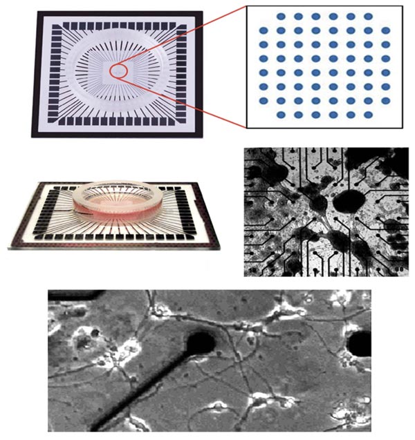
Fig. (2) Multi-electrode arrays (MEA). The image presents a diagram of MEAs (top row), a photograph of a MEA that more clearly depicts the culture dish situated above the electrode array (Left of middle row), and a micrograph of neuronal clusters within the MEA. The bottom panel presents a higher magnification image of neurons on a MEA; this image contains an electrode with its associated connecter and axons elaborated by neurons cultured on the MEA. MEAs are capable of recording the electrical activity of cultured neurons.