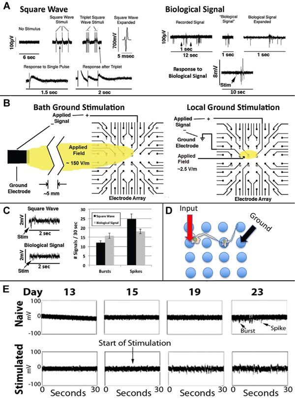
Fig. (8)
Panel A: Representative signals from an unstimulated mature network (“No stimulus”), single and triple biphasic square wave signals, the Biological Signal, and network responses to each. Panel B: Electric field generated using the bath ground electrode and that following localized stimulation. Panel C: Network responses following localized stimulation Panel D: Diagram of localized stimulation depicting how a neuron situated on the stimulated electrode could generate a downstream response of a second, synaptically connected neuron, which could be detected at an electrode in contact with the downstream neuron. Panel E: Network response following a single stimulation with the Biological Signal on day 15 after plating, which hastened the appearance of both spikes and bursts. Modified from [27].