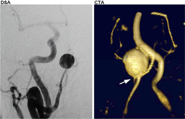
Fig. (2) Right posterior inferior cerebellar artery (PICA) aneurysm as imaged by DSA (left) and CTA (right). The right vertebral was not amenable to selective catheterization, and the subsequent innominate artery injection does not reveal much detail to the aneurysm and its vascular relationships compared with the result from the CTA. Note the relationship to the basilar artery, not seen with DSA, and PICA take-off at the base of the aneurysm (arrow) seen on CTA.