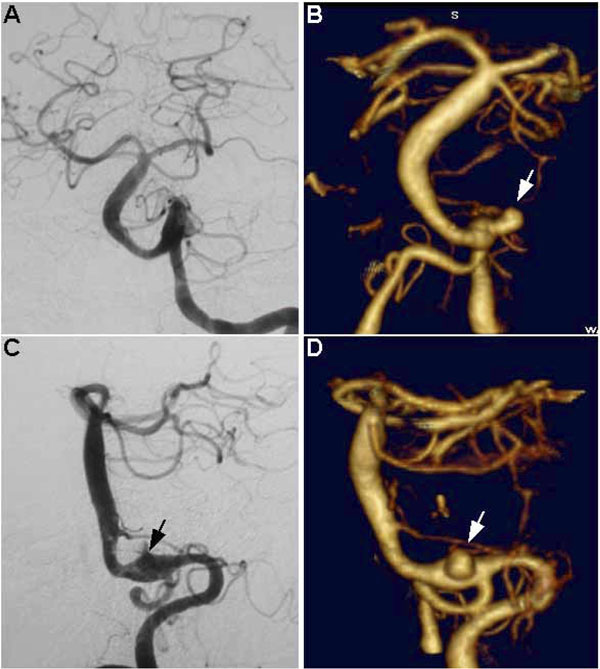
Fig. (5) Fusiform basilar artery aneurysm with a saccular aneurysm at the left vertebrobasilar junction. Note how this aneurysm is poorly seen on DSA (A and C, arrow shows probable location) and was initially missed using this modality, whereas it is unequivocally visualized on CTA (B and D, arrows). A and B represent anterior-posterior (AP) views, C and D represent lateral views.