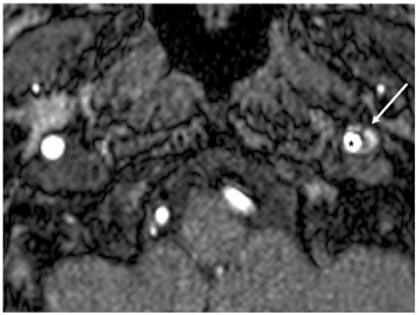
Fig. (1)
Axial magnetic resonance imaging scan showing a semilunar hyperintense signal due to the mural hematoma (white arrow) and an eccentric hypointense signal due to the residual lume, (black star).