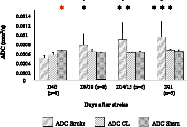
Fig. (3) ADC changes at different time points after stroke. Measures were collected in the stroke region (ADC), in the CL mirror region (ADC CL), and a correspondent region of a sham brain bilaterally (ADC sham). ADC was lower in strokes than in shams at D4/5 (p< 0.01, *) and ADC values at D4/5 were statistically different from values at D9/10 (p<0.05, *), D14/15 (p< 0.01, **) and D21 (p< 0.001, ***).