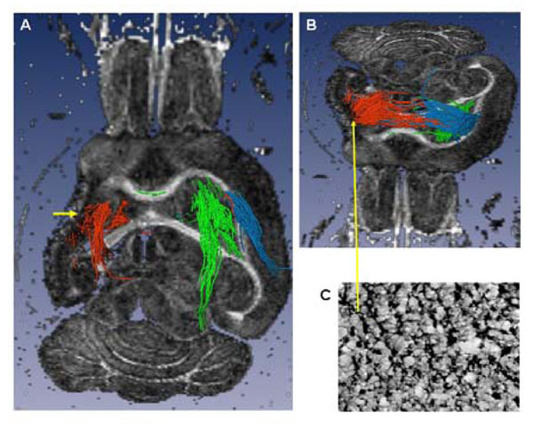
Fig. (6) Ex vivo tractography (A axial slice, ventral view and B tilted axial slice, dorsal view) of a brain with a large lesion involving the full anatomic extent (rostral to caudal) of the striatum and cortex, and the antero-lateral extent of the external capsule (EC). A) The lateral ventricle is enlarged, there is loss of striatal, cortical (cystic) and EC tissue (short yellow arrow): the peri-lesional trajectories (red) pass through the posterior, un-involved part of the EC and through the thalamus. In comparison, the contra-lesional trajectories (green) are similar to those seen in the sham brain, (Fig. 5D). B) The dorsal peri-lesional tracts continue to the CL EC. Tracts from the dorsal CL EC (blue) are similar to those originating from the peri-lesional region. C) Tissue region within the peri-lesional tissue showing increased GAP43 expression