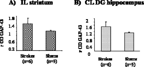
Fig. (7) A) GAP43 rOD between strokes and shams in the A) IL striatum and B) CL DG hippocampus. GAP 43 staining was much stronger in the stroke CL DG hippocampus of the stroke brains compared to the shams, where it is normally expressed at a low level. In the IL striatum the axon terminals are specifically stained for GAP 43 in the stroke mouse and not in the sham.