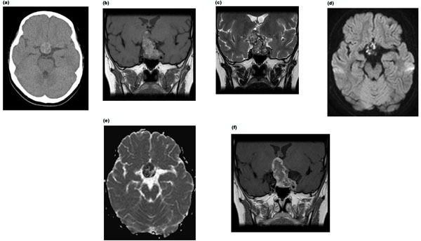
Fig. (1) Brain non-contrast CT shows a heterogeneous high-density mass in the suprasellar region (a). On MRI, T1-weighted coronal images show a lobulated mass with mixed high-intensity signal in the suprasellar region (b). T2-weighted coronal slice image shows a lobulated mass with mixed high-intensity and low-intensity signal in the suprasellar region (c). Diffusion-weighted image shows a heterogeneous high-intensity mass (d), and ADC map shows low-intensity mass. (e). Coronal TI-weighted gadolinium-enhanced MRI shows a mass with ring-like enhancement (f).