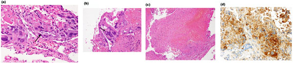
Fig. (2)
Photomicrographs of the biopsy specimens show mononuclear cytotrophoblast (arrow,
a
), a large multinuclear syncytiotrophoblast (arrow,
b
) with hemorrhage and necrosis (
c
). Immunohistochemical staining for hCG is strongly positive (
d
).