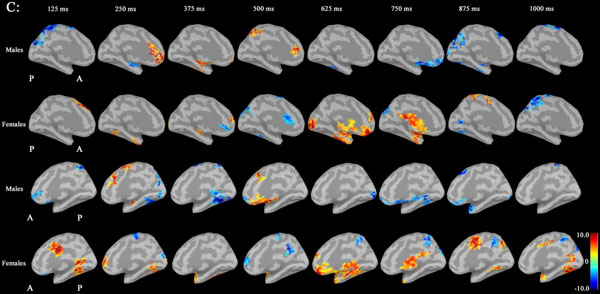
Fig. (1C)
Source maps of medial images in males and females at 100-150Hz: ERS in males was increased in the left rACC region at 250 ms, compared to females. ERD in females was increased in the left rACC region at 375 ms, compared to males. ERS in females was increased in both medial temporal regions at 625 ms, compared to males. (ERD: event-related desynchronization; ERS: event-related synchronization; rACC: rostral anterior cingulate cortex).