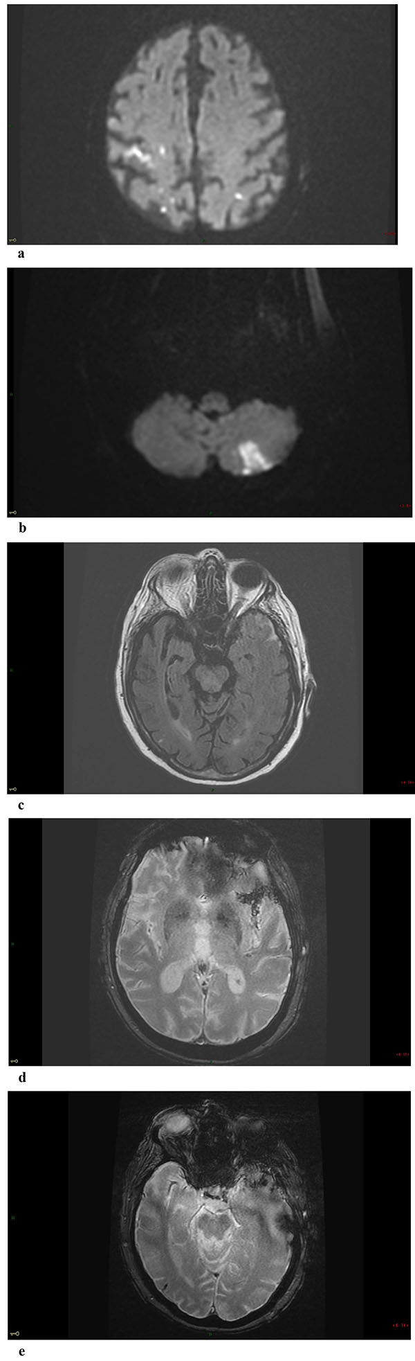
Fig. (1)
Axial DWI shows acute small embolic infarcts in territories of both middle cerebral arteries distal territory (a) and of the left postero-inferior cerebellar artery (a-b). Axial FLAIR, fluid attenuated inversion recovery (c) and axial T2 GRE images (d-e) show acute SAH filling partially the left sylvian fissure, the adjacent temporal convexity and a small sulcus at the right temporal convexity.