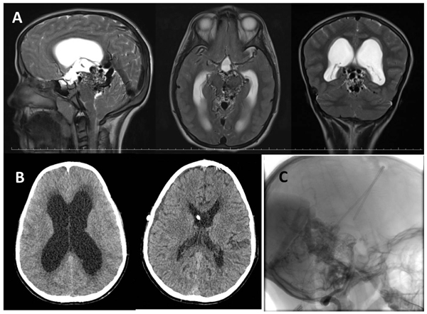
Fig. (1) A) T2 weighted MR scans showing a mesencephalic AVM that caused hydrocephalus with obstruction of aqueduct in sagittal, axial and coronary planes. B) A control CT showing the enlarged lateral ventricle with transependymal edema (left), and the collapsed ventricles following VP shunt procedure, with tip of the ventricular catheter (right). C) A DSA view perimesencephalic AVM in lateral plane, with intraventricular catheter.