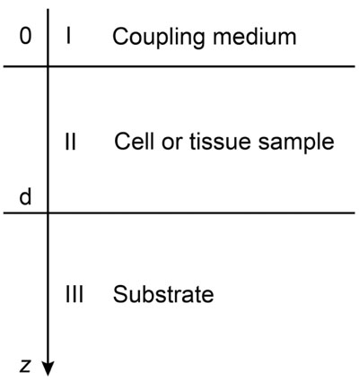
Fig. (4)
Layered model. The model depicts the cell/substrate structure. The cell or tissue samples are positioned between the coupling medium and the substrate. The coupling medium is typically a buffer solution with similar acoustic properties to the ones of distilled water at a given temperature.