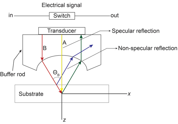
Fig. (6)
Schematic diagram for the V(z) calculations using the Ray theory method. The ultrasound transducer is positioned at the focus point above the substrate and the sample. The buffer rod is depicted in the above schematic. Examples of specular (yellow) and non-specular reflections (blue) are shown.