Fig. (7)
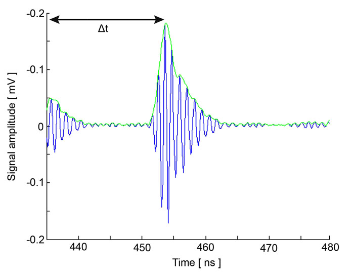
Quantitative analysis using time-resolved acoustic microscopy. Radio-frequency signal acquired during the scanning of MCF-7 cells. Echoes originating from the surface and the cell/substrate interface with the acoustic lens in focus are resolved on the time axis. The first echo received is due to reflections from the surface of the cell. The second echo is reflected off the cell/substrate interface. Note that the interface signal is much stronger compared to the one reflected from the cell surface.