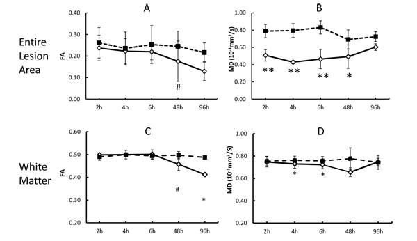
Fig. (3) Demonstration of temporal changes of FA and MD in entire lesion regions and white matter bundles within the lesion areas. A) Progressive changes of FA, and B) MD in the entire lesion regions after stroke insult. C) Progressive changes of FA, and D) MD in the white matter bundles after stroke insult. h: hours post stroke; solid line: stroke side; dash line: contralateral side; *, p<0.05, **, p<0.01, paired student’s t test between stroke side and contralateral side.