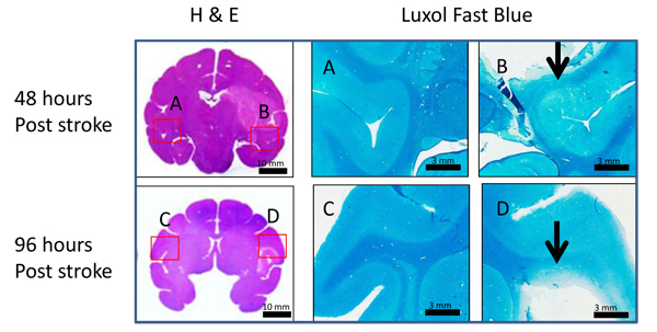
Fig. (5) Illustration of H & E and Luxol fast blue stained images. H & E (left column) and Luxol fast blue staining images (right column) in monkey brains 48 (top row) and 96 hours (bottom row) after MCA occlusion. Scale bars are shown at the right bottom of each picture. The corresponding regions of luxol fast blue staining pictures are labeled in the H&E staining whole brain pictures with A, B, C, and D (left: contralateral, right: ipsilateral) . Arrows point to abnormal white matter fibers.