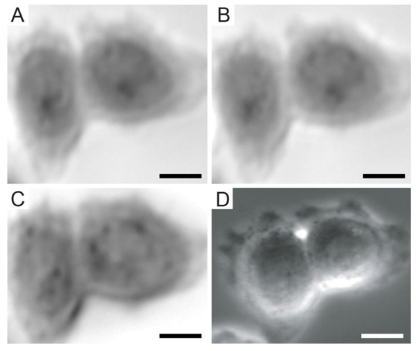
Fig. (3)
Acoustic and optical images of HeLa cells: (A) C-scan was taken in focus at z = 0; (B) C-scan of the same cells taken in focus 10 min after “A”; (C) acoustic image taken at defocus (z = 5 µm) (micrometers) 6 min after “A”; (D) brightfield micrograph taken prior the acoustic acquisition. The field of view of the acoustic images was 50×50 um. Magnification of the optical image was 40× (Figure from Weiss, Anastasiadis et al. © 2007 IEEE).