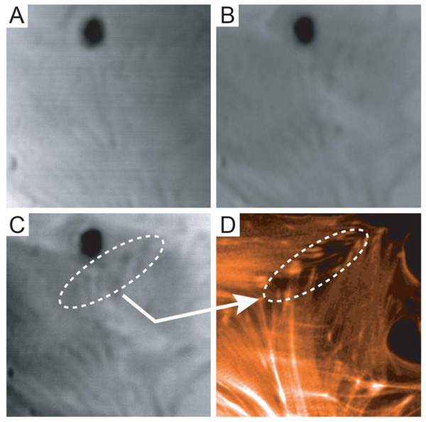
Fig. (8)
Acoustic and optical images of embryonic chicken heart muscle cells: (A) acoustic image taken at a defocus of z = - 4 µm; (B) acoustic image taken in focus (z = 0); (C) acoustic image at a defocus of z = 4 µm; (D) fluorescence image stained for F-actin. The field of view of the acoustic images is 50×50 µm (Figure from Weiss, Lemor et al. ©2007 Ultrasound in Med. & Biol.).