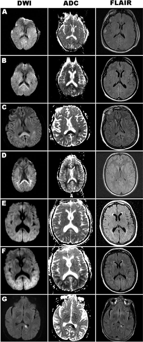
Fig. (1) Axial brain MRI images of DWI, ADC, and FLAIR sequences obtained in each case. A, Case 1, early MRI (Day 3) without splenium or cortical diffusion signal. B, Repeat MRI of case 1 (Day 10), with bilateral DWI-bright and ADC-dark splenium signal. C, Case 2 (Day 7), prominent DWI-bright and ADC-dark splenium signal, with subtle cortical ribbon signal, hyperintensity of these regions also present on FLAIR sequence. D, Case 3 (Day 8), DWI-bright and ADC-dark signal in the splenium and subcortical white matter, abnormal signal not well seen on FLAIR. E, Case 4, early MRI (Day 1) with minimal pathology. F, Case 4, late MRI (Day 6) with DWI-bright and ADC-dark signal in the splenium and occipital/parietal regions, with subtle corresponding hyperintensity signal on FLAIR. G, Case 5 (Day 3), isolated ischemic lesion in the left splenium, seen on all three sequences.

