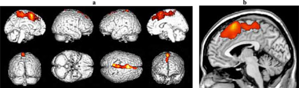
Fig. (2)
(a)
Three-dimensional standard rendering of the brain showing the areas of reduced cerebral blood flow in the disexecutive MCI group.
(b)
Sagittal section showing significant hypoperfusion in the anterior cingulate cortex.