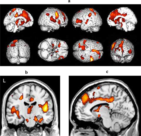
Fig. (3) (a) Three-dimensional standard rendering of the brain showing the areas of reduced cerebral blood flow in the multidomain MCI group. (b) Coronal section showing significant hypoperfusion in mediotemporal regions. (c) Sagittal section showing significant hypoperfusion in the anterior and posterior cingulate cortex.