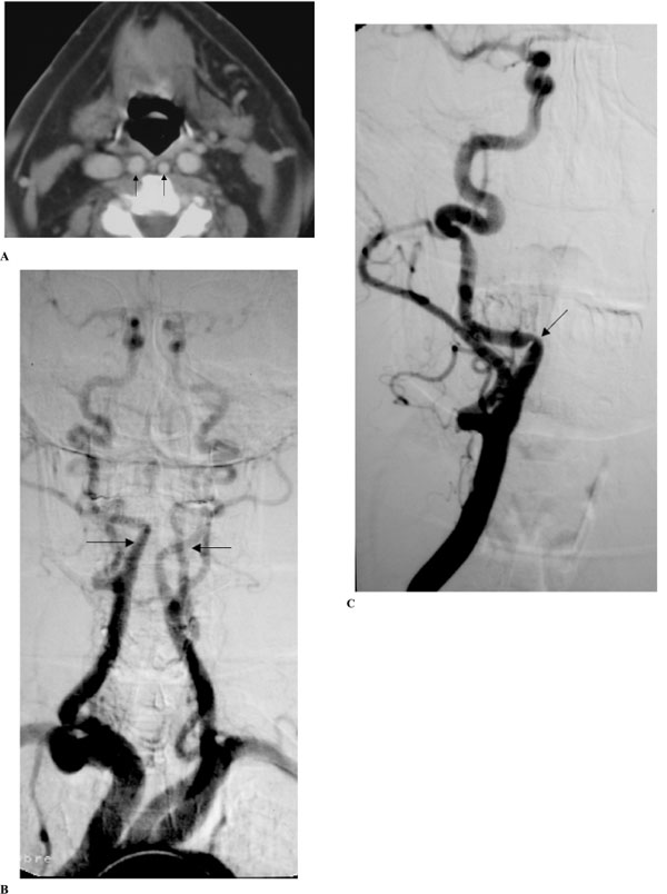
Fig. (3) (A-C). Extended bilateral retropharyngeal course of both internal carotid arteries (kissing carotids), (patient #12).
A: Axial contrast CT scan in early-enhanced phase throughout the oropharynx. Notice medialization of both ICA’s reaching the retropharyngeal midline (arrows), and also medialization of both internal jugular veins. B and C: Digital subtraction angiography in anteroposterior projection. Upper aortogram of both ICA’s in the early arteriographic phase (B) shows bilateral medialization of the cervical segment of the ICA’s (arrows). Notice tortuosity of the proximal portions of the right common and external carotid arteries. Selective injection of the right common carotid artery in early arteriographic phase (C) shows progressive medialization of the proximal segment of ICA and then abrupt change in the course of the artery and then the artery regains its normal position after a short horizontal segment. Notice extrinsic arterial notch pinpointing the place of arterial course change and producing a focal non-atherosclerotic stenosis (arrow). (Radiological classification of this patient was considered C,2.)