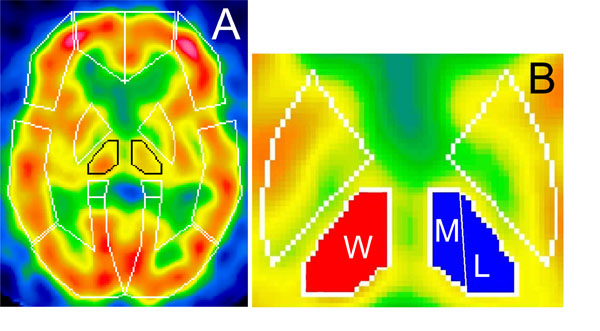
Fig. (1) Standardized brain SPECT images showing presets of ROIs template of 3DSRT (A) and subdivisions of thalamus used for CTUI measurements (B). ROI over thalami is outlined in black. Whole thalamus is marked as (W), medial and lateral parts as (M) and (L) respectively.