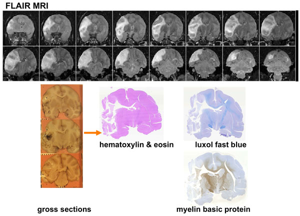
Fig. (5) Coronal images of the brain of a macaque that suffered a large stroke [11]. Top: In vivo FLAIR-T2 weighted MRI at day 10 showing herniation, mass effect and midline shift. Lower left: Gross coronal sections show tissue distortion, an obvious midline shift and some minor hemorrhage in the stroke core. Lower right: Hematoxylin and Eosin, luxol fast blue and myelin basic stained sections show a large area of necrotic tissue.