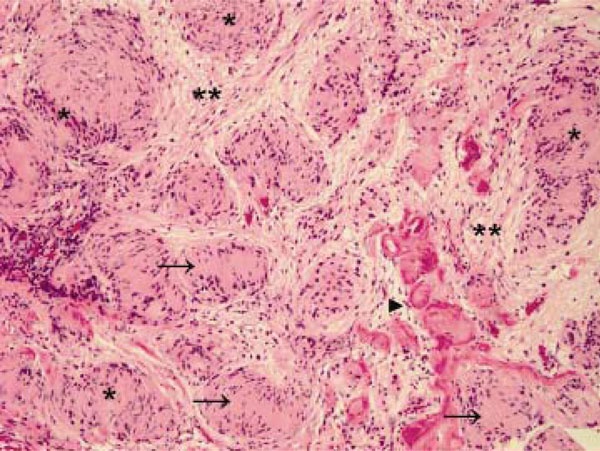
Fig. (2) Hematoxylin and eosin staining of the larger tumor as seen in Figure 1 after surgical removal. Hypercellular Antoni A areas (*), hypocellular Antoni B areas (**), Verocay bodies (arrows), and hyalinized blood vessels (arrowhead) are observed. Original magnification X 100.

