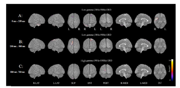
Fig. (1) MEG examination shows deviation of ERD power corresponding to target stimuli in the low (30-50 Hz) and high (50-100 Hz) gamma bands in patients with schizophrenia compared to normal controls (A paired test, FWE p < 0.005).A: Low gamma ERD (30-50 Hz) power is significantly decreased in the right inferior temporal and a small area on the left of the midline frontal lobes at 0-250 ms.B: Low gamma ERD power is significantly decreased in the left midline dorsal frontal area, a bilateral small midline parietofrontal area, and a small area of the left upper temporal lobes at 250-500 ms.C: High gamma ERD (50-100 Hz) power was significantly decreased in a small area of the left occipital lobe at 500-750 ms.