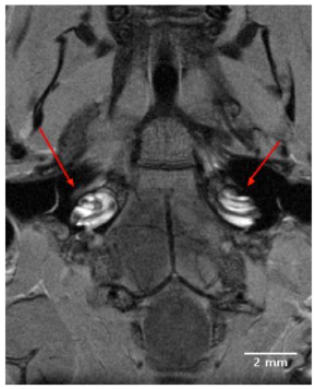
Fig. (1) A 9T, 2D transverse orienting slice showing the bilateral anatomical organization of the normal mouse external auditory meatus, middle ear and cochlea located in close proximity to the ipsilateral and contralateral brainstem. The scala tympani and scala vestibuli of the cochleae are clearly enhanced for visualization by the IV injected Gd complex, in contrast to the unenhanced dark rings of the scala media.