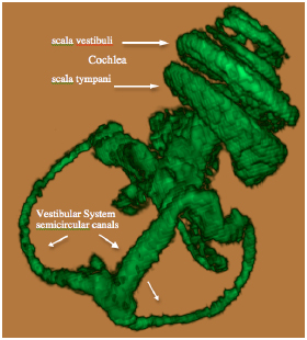
Fig. (2B)
3D volume rendered MRI at 9.4 T of the normal mouse vestibulo-cochlea labyrinth revealing the superior, inferior and lateral semicircular canals, utricle, sacculus, and the scala tympani and scala vestibuli in each turn of the cochlea.