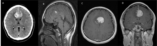
Fig. (1A)
Axial computed tomography demonstrates an acute inter-hemispheric haemorrhage.
B
) Sagittal T1-weighted sequence.
C
) Axial post-gadolinium T1-weighted sequence.
D
) Coronal post-gadolinium T1-weighted sequence, indicating tumour location.