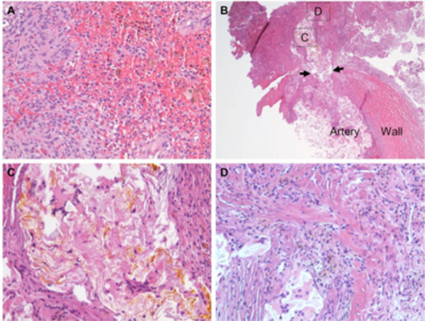
Fig. (3) Micro-photographs showing histopathologic findings. A) A meningioma with meningothelial features, fresh haemorrhage and
hemosiderin deposition (brown colour, suggestive of previous haemorrhage). B) An aneurysm (arrows pointing to the neck) at the wall of a
muscular artery. C) The site specified in B) showing aneurysm embolisation N-butyl-2-cyanoacrylate material (yellow colour) and
surrounding reactive changes, including multi-nucleated giant cells. D) The site specified in B) showing aneurysm rupture with surrounding
hemosiderin deposition. Original magnification 200×, A, C and D; 25×, B.