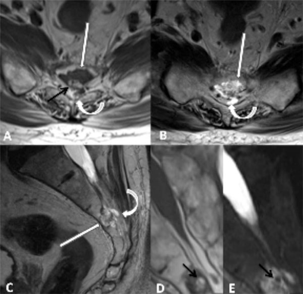
Fig. (1) Axial T1-weighted (A), axial T2- weighted (B), and sagittal T2-weighted (C) images demonstrate a lobulated mass involving S3
and the dorsal superior body of L4 (straight white arrows) with heterogeneous T1- and T2 signal with areas of intermediate and high signal.
There is subtle focal dorsal extraosseous extension of the tumor into the sacral canal (curved arrows). Subtle marrow edema along the ventral
and superior aspect of the tumor margin is noted on sagittal T2-weighted image (C). Magnified sagittal T1- (D) and fat suppressed sagittal
T2-weighted (E) images as well as axial T1- weighted image (A) demonstrate lobulated foci of intralesional fat (black arrows) with complete
suppression of fat signal on fat suppressed T2-weighted images (E).