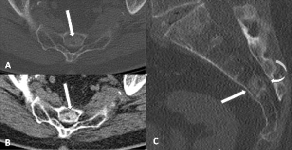
Fig. (2) CT images of the sacrum with axial bone (A) and soft tissue (B) algorithm and sagittal bone (C) algorithm demonstrate a sclerotic
lesion within S3 and the superior body of S4 (white arrows), which is diffusely sclerotic with a relatively narrow zone of transition.
However, there is subtle focal cortical permeation involving the cortex along the dorsal aspect of the lesion, with subtle extraosseous
extension into the sacral canal (curved arrow).