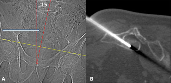
Fig. (3) Scout image (A) with patient prone demonstrates oblique positioning with spine and pelvis positioned approximately 15 degrees to
the right (red angle). This aligned the left S4 pedicle with the scanner gantry (white arrow.) Procedural CT image (B) demonstrates an 11-
gauge needle extending through the left S4 pedicle into the intraosseous tumor at S3.