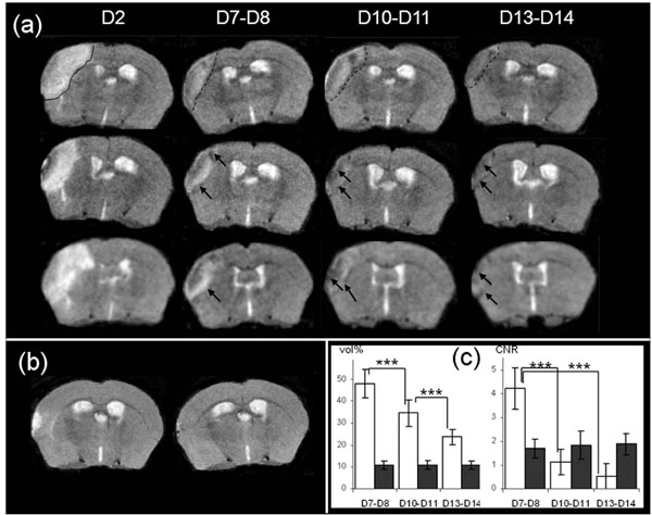
Fig. (3) (a): Brain T2-weighted MR images from 3 mice at Days 2, D7-8, D10-11 and D13-14 after MCAo. Edema is visible at Day 2
(delineated by a continuous line, upper row image). The infarct core is delineated by a dotted-line at D7-D8, D10-D11 and D13-D14 in the
same mouse brain (upper row), and low intensity area (LA) designated by an arrow in the brain of the other mice (b): Brain T2-weighted MR
images from 1 representative sham mouse at Days 2 and 7 after procedure (c): Evolution of apparent volume and CNR of infarct core (in
white) and of low intensity area (in black) at Days 7-8 (n=25), Days 10-11 (n=16) and Days13-14 (n=16) after MCAo. Volumes are
represented as a % of the hyperintensity area volume delineated at Day 2. The differences in the mean values of infarct core volume against
time were statistically significant (DFr=30; F=126.455; p<0.001), and p<0.001 for all pairwises. For LA volume there was not a statistically
significant difference (DFr=30; F=0.268; p=0.757). The CNR were calculated as described in the Material and Methods section. The
differences in the mean values of infarct core CNR against time were significantly different (DFr=30; F=251.219; p<0.001), and p<0.001
between D7-D8 and D10-D11 or D13-D14), and there was not a statistically significant difference for LA CNR (DFr=30; F=1.550;
p=0.229).