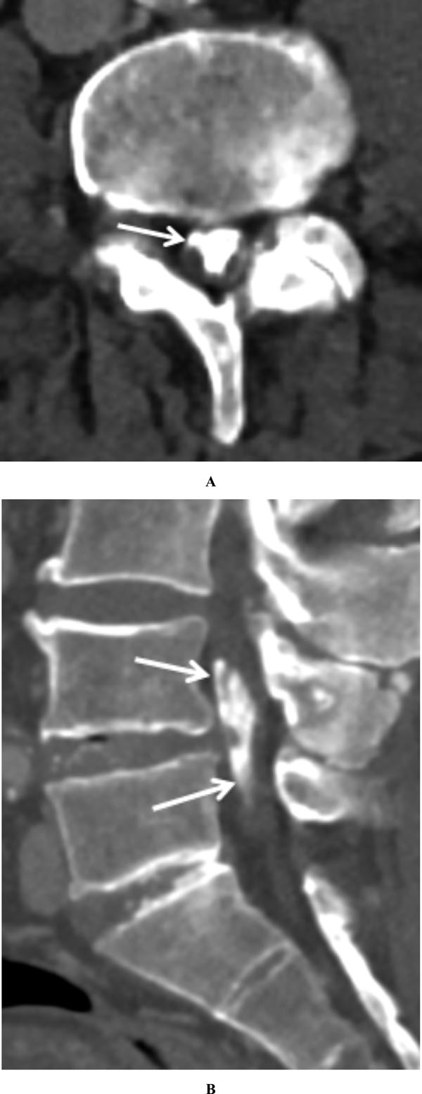
Fig. (4) Axial CT image through the lower lumbar spine (A) demonstrates central ossification with the thecal sac (arrow), corresponding to
regions of T2 hypointensity on MRI (Fig. 3A). Sagittal reformatted image (B) demonstrates the craniocaudal extent of the arachnoid ossification
(arrows). Multilevel degenerative changes are partially visualized.