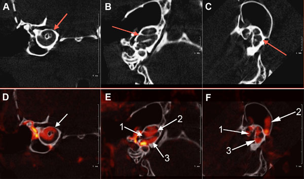
Fig. (3) Axial view of the μCT scans of the osseous mouse cochlea chambers (white) including (arrows) the scala tympani area (A), scala
media, scala vestibuli, vestibular apparatus, and semicircular canals (B) and (C). Fig. (3): (D). white arrow shows μCT fused with a CT
segmentation of the perilymphatic space (red) sections of the mid-cochlea; (E). (1) Scala media; (2) scala vestibuli and Scala tympani (3) (F).
Contiguous osseous labyrinth of the mouse inner ear showing the (1) scala vestibule, (2) semicircular canals of the vestibular apparatus, and
(3) scala tympani of the cochlea.