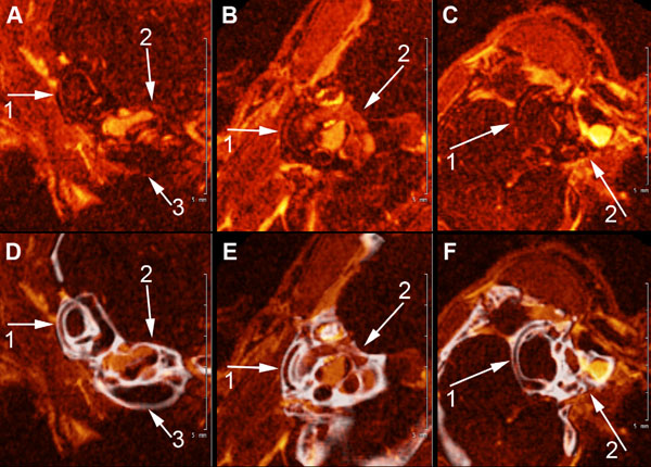
Fig. (4) A, B, and C show high field MRI Gd-enhanced images of the scala tympani, scala vestibuli and semi-circular canals of the mouse
inner ear (1) semicircular canals of the vestibular apparatus, (2), scala vestibuli and scala tympani (3). 4. D, E, F show fusion images (in
white) of CT segmentation of the cochlea and semicircular canals of the contiguous osseous labyrinth of the mouse inner shown in A, B, and
C.