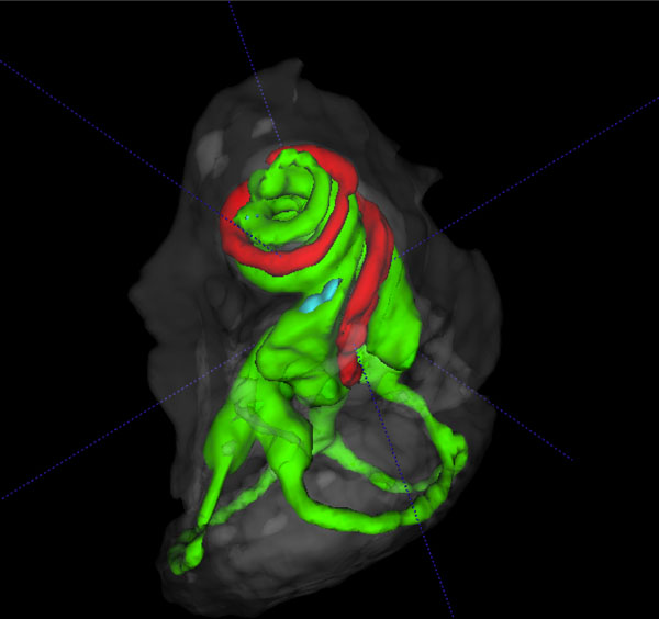
Fig. (5A) 3D volume rendered cochlear and vestibular apparatus of
the mouse inner ear with focus on the cochlea, showing the contrast
enhanced perilymphatic spaces of the scala tympani (Green), scala
vestibule (Green) and the scala media (red), and the lateral,
posterior and anterior semicircular canals (Green), surrounded by
the osseous labyrinth (White). The vestibular system is seen below
the cochlea.