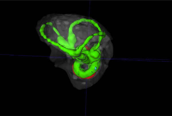
Fig. (5B) Reverse view of a 3D volume rendered vestibular
apparatus and cochlea of the mouse inner ear with focus on the
vestibular system, showing the contrast enhanced perilymphatic
spaces of the lateral, posterior and anterior semicircular canals
(Green), surrounded by the osseous labyrinth (White). The cochlea
scalae are seen below.