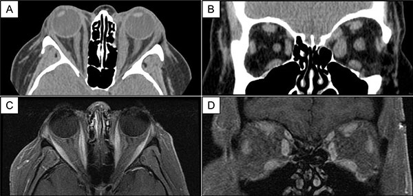
Fig. (1) Case 1. Axial (A) and coronal views (B) of CT imaging of the orbits show enlargement of multiple extraocular muscles, left greater than right, with tendon involvement. Eleven months post-diagnosis, axial (C) and coronal views (D) of T1-weighted, fat-suppressed MRI of the orbits demonstrate reduced extraocular muscle caliber with steroid-sparing immunosuppression.