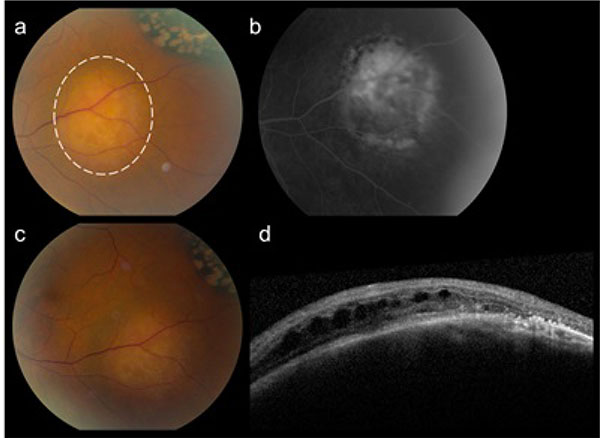
Fig. (2) The inferotemporal choroidal carcinoid metastasis in the left eye. Fundus photograph at presentation (a, dotted line). The hyper/hypopigmented area superiorly of the choroidal metastasis in (a) represents a congenital hypertrophy of the retinal pigment epithelium (CHRPE) with lacunae. Pre-treatment fluorescein angiography 1.5 months after presentation (b). The choroidal carcinoid lesion shows chronic surface changes but remains status quo after 10 cycli of Sandostatin®(c). OCT scan demonstrates intraretinal fluid overlying the lesion (d).