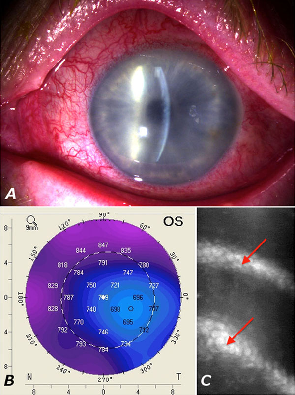
Fig. (2) (A): Slitlamp photograph showing conjunctival injection and corneal oedema of the left eye.(B): Pentacam topography demonstrating an increased corneal thickness of 750 μm in the center of the cornea, and 847 μm in the periphery.(C): Confocal microscopy of the corneal endothelium. The image is blurred due to the oedema. The arrow indicates folds in Descemets membrane. Corneal endothelial cell density measurement was not possible due to the oedema.