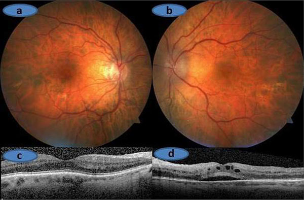
Fig. (1)
(A)
and
(C).
Right Eye, Normal optic disc with normal macula scan.
(B)
and
(D).
Left eye, Swollen optic disc and subfoveal subretinal + intraretinal fluid on the OCT scan.