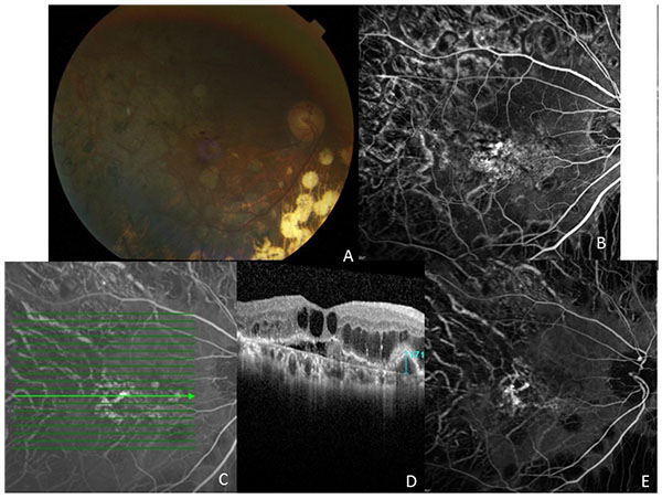
Fig. (1)
(A) Fundus photograph of the right eye of patient 1. Note the small subretinal hemorrhage within the macula and peripheral panretinal laser photocoagulation for proliferative diabetic retinopathy. (B) Fluorescein angiography shows leakage in the inferotemporal fovea. (C, D) ICG angiogram showing PCV complex with nasal polyp and horizontal line showing ICG location for optical coherence tomography line scan. Note the characteristic inverted U-shaped polyp with a height of 171 um, the adjacent branching vascular network (shallow elevation of the RPE), and diffuse intraretinal edema with serous detachment. (E) ICG angiography shows multiple hyperfluorescent polyps with associated branching vascular network.