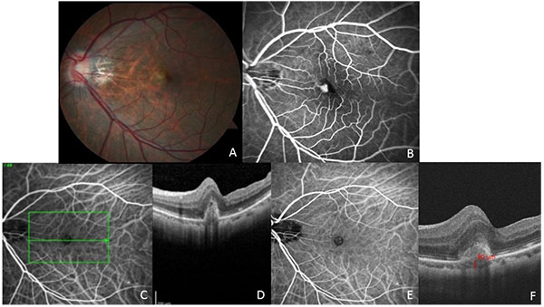
Fig. (2)
(A) Fundus photograph of the left eye of patient 2 shows nasal retinal thickening. (B) Fluorescein angiography shows leakage in the area of nasal retinal thickening (C, D) ICG angiogram showing central hyperfluorescent lesion with hypofluorescent border and a horizontal line showing ICG location for optical coherence tomography line scan. Note the characteristic inverted U-shaped polyp overlying macular edema and cystic change. (E) Indocyanine green angiography shows a hyperfluorescent polyp with a surrounding hypofluorescent halo. (F) Choroidal thickness measurement on OCT confirming thin choroid (80 microns) underneath the fovea.