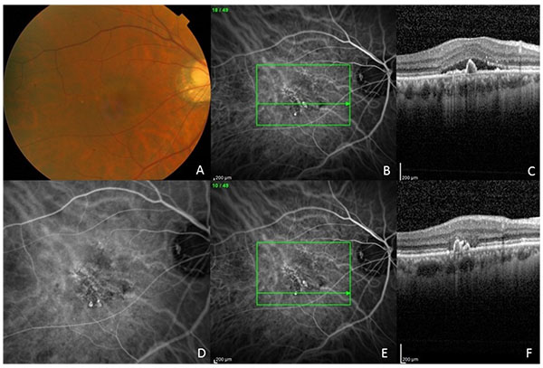
Fig. (3)
(A) Fundus photograph of the right eye of patient 3. Note the myopic fundus with peripapillary RPE atrophy and laquer cracks and serous macular detachment. (B, C) ICG angiography showing PCV complex with polypoidal dilations and branching vascular network. Note the inverted U-shaped polyp and adjacent branching vascular network with shallow elevation of the RPE. There is overlying serous detachment. (D) ICG angiogram showing magnified view of PCV complex with branching vascular network and terminal polyps. (E, F) ICG angiogram of PCV complex with horizontal line scan through polyps inferiorly outside the area of leakage. Note the multiple inverted U-shaped polyps and loss of the photoreceptor layer above the polyps.